
Snapshots of Synapses „in Action“
A study published online at Neuron on 28 September presents a novel method to visualize changes in the nanoscale organisation of stimulated synapses in brain tissue by electron microscopy.
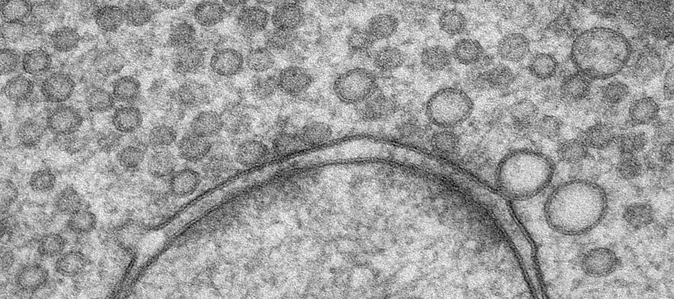
© MPI f. experimentelle Medizin / Benjamin Cooper & Cordelia Imig
Nerve cells communicate at synapses, highly specialized contact points that represent the basic signalling units of the brain. Synaptic signalling takes place within milliseconds, and its efficacy and fidelity determines many behavioural and memory processes. However, experimentally capturing the ultrastructural basis of synaptic activity, such as synaptic vesicle fusion or endocytic membrane recycling, has remained extremely challenging.
Cordelia Imig and her colleagues Nils Brose and Benjamin Cooper at the MPI of Experimental Medicine (Germany) have now published a methodological approach for functional electron microscopy to be applied to cultured brain tissue. This became possible by combining optogenetics, which enables the activation of distinct neuron types within neuronal networks by short blue light pulses, and rapid cryofixation of cultured brain tissue within milliseconds after light stimulation (“flash-and-freeze”). To demonstrate the power of their approach, the technique was used to study highly specialized hippocampal mossy fiber synapses, which form huge synaptic terminals, can support neurotransmitter release reliably for long periods of activity, and are critically important for spatial memory formation. Combining the approach with high-resolution 3D electron microscopic imaging revealed that in this synapse, strong stimulation results in a depletion of vesicles and compensatory membrane recycling near neurotransmitter release sites.
In the future, this technique will make it possible to dissect the molecular control of distinct activity-induced synaptic membrane trafficking steps in neurons embedded within networks. The results from corresponding studies will contribute to a better understanding of the molecular mechanisms of fundamental synaptic processes that ultimately determine synapse function and pathological dysfunction.
Reference
Imig C, López-Murcia FJ, Garcia-Plaza IH, Mortensen LS, Schwark M, Angibaud J, Nägerl V, Taschenberger H, Brose N & Cooper BH (2020) Ultrastructural Imaging of Activity-Dependent Synaptic Membrane Trafficking Events in Cultured Brain Slices. Neuron; DOI:https://doi.org/10.1016/j.neuron.2020.09.004